Quick answers
The questions patients actually ask, answered briefly. Each one links to the section that covers it properly — use this as the page's contents list.
One side of my nose has been blocked for weeks, sometimes with a little blood — can I wait and watch?
No — symptoms that stay on one side, particularly blockage with blood-stained discharge, facial numbness or eye symptoms, should be examined rather than treated blindly with sprays. This is uncommonly something serious, but it is the one pattern where an ENT examination should not be postponed.
Does nasal endoscopy hurt? — Naak ki doorbeen se dekhne mein dard hota hai kya?
It is usually described as odd and briefly uncomfortable rather than painful — a pressure or watering-eyes sensation for a few seconds as the scope passes. Trials of numbing sprays have not shown a clear reduction in pain.
Read: what it feels like, and the evidence on numbing spray →
Will I be given anaesthesia or put to sleep for it?
No — diagnostic nasal endoscopy is an outpatient examination done while you sit awake in the chair, with at most a spray in the nose. It usually takes a couple of minutes per side.
Why can't the doctor just look inside with a torch?
A torch and speculum show only the front of the nasal cavity, while most sinus drainage pathways sit several centimetres further back. The scope carries its own light and lens to that area.
Can nasal endoscopy tell me whether I have sinusitis?
It can show the specific signs that guidelines ask for — polyps, pus from the middle meatus, or swelling there — which, alongside your symptom history, is what confirms chronic rhinosinusitis. It cannot grade disease inside the sinus cavities themselves.
My nose blocks up because of allergy — do I still need a scope?
Not always; a clear allergy pattern with a good response to treatment often needs no endoscopy at all. It becomes useful when symptoms persist despite treatment, or when the picture does not fit allergy alone.
Will I need a CT scan as well?
Often not — endoscopy and CT answer overlapping questions, and imaging is reserved for specific reasons such as planning surgery or an unclear or one-sided finding. CT is not performed at the clinic and would be arranged separately.
My child has been told to have a nasal endoscopy — is that reasonable?
In cooperative children a thin paediatric scope is generally well tolerated and gives a direct view of the adenoid, which is otherwise judged indirectly. Whether it is attempted depends far more on the child than on the equipment.
Can I drive home and go back to work afterwards?
Most people return to normal activity immediately, since nothing sedating is given for a routine diagnostic examination. A brief nosebleed or a few sneezes settle within minutes.
Why a scope, when the nose is right there
The part of the nose that causes most trouble is the part you cannot see from the front. A head-light and a nasal speculum give a reasonable view of the septum, the front of the inferior turbinate and the nasal vestibule — and that is genuinely useful, since a deviated septum or a swollen front turbinate explains a great many blocked noses. But the sinuses do not open at the front. They drain through narrow channels several centimetres back, in a region called the middle meatus, tucked under the middle turbinate.
A nasal endoscope is a thin telescope, roughly the width of a thick pen refill in adults and considerably thinner in children, that carries a light source and a lens to that area. The image appears on a monitor at magnification, so mucosal swelling, a strand of pus, a small polyp or an area of crusting are seen directly rather than inferred.
It is an examination, not an intervention
It is worth separating two things that share a name. Diagnostic nasal endoscopy is what this article describes: an outpatient look, performed awake, taking a few minutes. Endoscopic sinus surgery uses the same optical principle but is an operation performed under anaesthesia in a theatre — at an attached hospital, not at the Golf City clinic — and is a separate decision made only after medical treatment has been given a fair trial.
Which leads to the more useful question: once the scope is in, what is actually being looked for?
What the view actually shows
Endoscopy is looking for a small number of specific findings, not browsing. In chronic rhinosinusitis, the European Position Paper on Rhinosinusitis and Nasal Polyps (EPOS 2020) defines the condition as the right combination of symptoms lasting long enough, plus either endoscopic signs or CT changes. The endoscopic signs it names are narrow and concrete: nasal polyps, mucopurulent discharge coming primarily from the middle meatus, or oedema and mucosal obstruction primarily in the middle meatus.
That structure matters for a patient, because it explains why an ENT may seem uninterested in a long description of symptoms alone. Symptoms establish the possibility; the examination is what confirms or removes it. The 2025 update of the AAO-HNSF adult sinusitis guideline makes the same point from the other direction, recommending that a clinical diagnosis of chronic rhinosinusitis be confirmed with objective documentation of sinonasal inflammation — by anterior rhinoscopy, nasal endoscopy or CT.
Beyond the sinus question
- Structural causes of blockage — septal deviation and spurs further back than a speculum reaches, turbinate enlargement, adhesions from previous surgery or injury, a septal perforation.
- The nasopharynx — the space behind the nose, including the adenoid pad and the Eustachian tube openings, which is relevant to persistent ear blockage and to mouth breathing in children.
- A bleeding point — in recurrent nosebleeds, a vessel too far back for a front-of-nose examination to find.
- Anything one-sided — a mass, unusual crusting or a polyp confined to one side, which is treated as a finding needing explanation rather than reassurance.
Key limitation: endoscopy shows the surfaces the scope can reach. It does not see inside the maxillary or frontal sinus cavities, cannot measure how much disease sits within them, and does not by itself distinguish a benign polyp from a growth that needs tissue diagnosis. A normal endoscopy also does not rule out every cause of a blocked nose — non-allergic rhinitis, medication overuse and some structural problems can coexist with an unremarkable view.
When it changes the plan — and when it doesn't
The honest answer is that many people with nasal symptoms never need a scope. A first episode of acute sinusitis, a clear seasonal allergy pattern, a simple viral cold — none of these are endoscopy questions, and a course of appropriate treatment answers them faster than a camera does. Endoscopy earns its place when the answer will change what happens next.
The situations where it usually does
- Symptoms lasting beyond about twelve weeks despite reasonable treatment, where a diagnosis of chronic rhinosinusitis needs to be confirmed rather than assumed.
- Suspected nasal polyps, where confirming their presence or absence changes both the medical plan and the discussion about surgery.
- Persistent one-sided symptoms of any kind — this is the strongest single indication, and the reason is covered further down.
- Recurrent nosebleeds where the source has not been identified at the front of the nose.
- Persistent smell loss that has not recovered, where the olfactory cleft needs to be inspected.
- Blockage that has not responded to a properly used nasal steroid spray, where the question becomes whether the obstruction is inflammatory or structural.
- Persistent ear fullness or one-sided fluid behind the eardrum in an adult, where the nasopharynx needs to be seen.
Equally, there are times to say no. Endoscopy in the middle of an acute infection is often uninformative and more uncomfortable than usual, and is generally better deferred unless there is concern about a complication. And a scope done because it is available, rather than because a question needs answering, adds cost and discomfort without adding clarity. That said, the most common reason patients decline is not cost — it is the expectation that it will hurt.
What actually happens, step by step
The whole thing is shorter than the consultation that precedes it. You sit upright in the examination chair, head supported, and the sequence runs roughly as follows.
1. The conversation and consent
The clinician explains what is about to happen and asks about anything that changes the approach: previous nose surgery, a known bleeding tendency or blood-thinning medication, recent nasal trauma, or a very narrow nose. Verbal consent is standard for this kind of outpatient examination.
2. Preparing the nose
A decongestant spray, sometimes combined with a local anaesthetic, may be used to shrink the lining and widen the passage. It takes a few minutes to work and tastes bitter as it trickles backwards. This step is optional rather than automatic, for reasons explained in the next section, and you should be asked which side of your nose feels more open.
3. The examination
The scope is lubricated and passed gently along the floor of the nose, then withdrawn and passed along a higher route, so that the different levels of the nasal cavity are inspected in turn. If the nasopharynx and larynx also need to be seen, the scope continues past the back of the nose and you may be asked to sniff, swallow, poke your tongue out or say "eeee" — each of these opens up an area that is otherwise hidden. You can breathe normally throughout; the scope occupies a small part of one nostril, not your airway.
4. Finishing
Both sides are usually examined unless one is clearly the problem. Images are captured for the record so that the appearance can be compared at a future visit. Total scope time is typically two to five minutes.
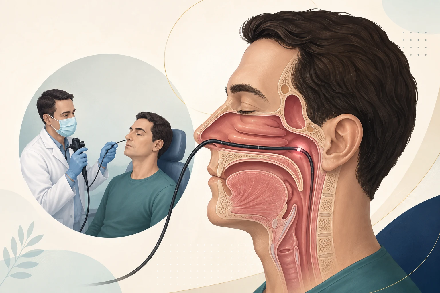
Nothing in that sequence involves cutting or injections. The part patients dread is the passing of the scope itself — which turns out to be the part the evidence has most to say about.
Does it hurt? What the evidence says about numbing spray
Most people find it strange rather than painful. The usual descriptions are a pressure sensation, a tickle that makes the eyes water, and an urge to sneeze — all lasting seconds. Discomfort is greater in a very narrow or very inflamed nose, and greater still if the scope is forced, which is why an unhurried technique matters more than any spray.
The spray question is genuinely interesting, because the intuitive answer is wrong. A Cochrane review of topical preparations before flexible nasal endoscopy found no clear evidence that they reduce pain or discomfort, and noted that some authors argue against routine use because of cost and unpleasant side effects. A separate systematic review and meta-analysis of eight randomised trials in 818 patients reached a similar conclusion: no difference in pain scores between local anaesthetic, vasoconstrictor and lubricating agents, with co-phenylcaine associated with greater taste unpleasantness and lidocaine, in one trial, with more pain. Both reviews are cautious about their own conclusions, since several of the underlying trials were small.
What that means in practice
It does not mean sprays are useless. A decongestant can widen a genuinely narrow passage and improve the view, and a single trial in that meta-analysis did find lower overall unpleasantness with xylometazoline. It does mean that being sprayed is not the thing standing between you and a comfortable examination, and that declining the anaesthetic — or being told it is not needed — is a defensible clinical position rather than a shortcut.
Complications are uncommon. The recognised ones are sneezing, a small amount of bleeding from mucosal contact, gagging when the scope passes further back, and occasionally light-headedness. A brief nosebleed is the one people notice, and adequate decongestion and gentle technique are what prevent it.
Nasal endoscopy in children
Children tolerate it better than most parents expect, but the decision is more finely balanced than in adults. Paediatric scopes are considerably thinner — down to around 2 mm — and in a cooperative, prepared child the examination is brief. In a distressed toddler it is neither kind nor informative, and there is no merit in forcing it.
The commonest reason to consider it is the adenoid. A child with persistent mouth breathing, snoring, a permanently blocked nose or recurrent ear fluid is often being assessed for adenoid enlargement, and endoscopy gives a direct view of the pad and how much of the airway it occupies — where a lateral X-ray gives a two-dimensional shadow and clinical examination alone is unreliable. Endoscopy is generally regarded as the reference standard for this assessment, with radiography as a practical alternative when a child cannot cooperate.
What makes it go well
- Explaining honestly beforehand: it feels strange, it is quick, it does not involve a needle.
- The child sitting on a parent's lap for younger ages, with gentle head support.
- Accepting a partial examination. A brief look at the nasopharynx often answers the question; a complete survey is not always necessary.
- Stopping if the child is genuinely distressed and reconsidering, rather than persisting.
If you are weighing this up for a child, the clinic's guides on persistent mouth breathing in children and when a child should see an ENT cover the surrounding decisions. What happens after the scope, for adults and children alike, is usually the part that gets least explanation.
Afterwards, and reading your report
There is no recovery period to speak of. Nothing sedating is given for a routine diagnostic examination, so driving, working and eating are unaffected — the exception being that if a local anaesthetic spray was used and your throat feels numb, it is sensible to wait around an hour before eating or drinking so you do not scald or choke yourself without noticing. Sneezing and eye-watering settle within minutes. A small amount of blood-tinged mucus for a nose-blow or two is common and not a cause for concern; bleeding that continues beyond a few minutes is worth reporting.
What a useful report contains
Findings are more helpful when they are specific. A report or a verbal explanation should tell you which side, which structure, and what the appearance means for the plan — not simply "endoscopy done, sinusitis present". Reasonable things to ask before you leave:
- What did you see, and on which side?
- Does it explain my main symptom, or only part of it?
- Has anything been ruled out by this examination — and what has not?
- Does this change the treatment, or confirm the treatment I am already on?
- Do I need any further test, and what question would that test answer?
That last question is the one that keeps investigations proportionate, because endoscopy sits alongside other tests rather than replacing them.
What endoscopy does not replace
Endoscopy and CT agree more often than patients assume. A systematic review of sixteen studies cited in EPOS 2020 reported a high correlation between endoscopy and paranasal sinus CT for the diagnosis of chronic rhinosinusitis (r = 0.85, 95% CI 0.78–0.94), which is part of why guidelines accept either as objective confirmation. Agreement is not equivalence, though, and the two answer different questions.
| Assessment | Main question it answers | What it cannot do alone |
|---|---|---|
| Nasal endoscopy | Are there visible signs of inflammation, polyps, obstruction or a one-sided abnormality in the reachable nasal cavity and nasopharynx? | Cannot see inside the sinus cavities or grade disease within them; cannot give a tissue diagnosis. |
| CT of the sinuses | How much disease is present within the sinuses, and what is the anatomy before surgery? | Involves radiation, is not needed for most patients, and can show incidental changes in people without symptoms. |
| Skin prick testing | Which inhaled allergens produce a reaction, to be interpreted against the history. | Does not show what the nose looks like or exclude a structural cause. |
| Biopsy | What a tissue abnormality actually is. | Only relevant when something has been seen that needs explaining; arranged separately when indicated. |
| Laryngoscopy | The appearance of the throat and voice box, often in the same sitting when symptoms include hoarseness or throat clearing. | Does not assess the sinuses. |
Where allergy is the driver, the nose may look inflamed on the scope without endoscopy telling you what it is reacting to. Skin prick testing answers a different part of the question, and for confirmed allergic rhinitis sublingual immunotherapy is a longer-term option matched to those results. If sinus symptoms and allergy symptoms have blurred together, the guide on seasonal allergies versus sinusitis is the better starting point.
Symptoms that should not wait for a routine appointment
- Persistent or progressive blockage of one nostril
- Blood-stained discharge or repeated nosebleeds from the same side
- Cheek numbness or facial swelling, loosening teeth, or suddenly ill-fitting dentures
- Eye symptoms such as bulging, double or reduced vision, redness, or swelling
- Swelling at the inner corner of the eye or persistent watering of one eye
- Severe headache with frontal swelling, confusion, neck stiffness, or a new neurological symptom
- A persistent foul smell only you can perceive, or one-sided crusting
Read the short guide to nasal symptoms that need prompt assessment →
The bottom line
Nasal endoscopy is a short outpatient examination that answers a specific question: what does the part of the nose you cannot otherwise see actually look like? It confirms or removes a diagnosis of chronic rhinosinusitis, finds polyps, locates a bleeding point, shows the adenoid in a child, and — most importantly — investigates anything happening on only one side.
It is not a routine add-on to every consultation, it is not a substitute for a CT scan when imaging is genuinely needed, and it is not a treatment. Its value comes from the question it is asked to answer and from a clinician who connects the view with your symptoms, your examination and your response to treatment so far.
HealthNest Clinic provides ENT consultation with nasal endoscopy, laryngoscopy, allergy skin prick testing and hearing assessment when clinically indicated, at Golf City, Lucknow.
Message on WhatsAppPractical questions
Logistics and edge cases that do not need a full section.
What should I bring to the appointment?
Bring a written list of your current medicines including any nasal sprays and their dose and frequency, plus any previous ENT reports, allergy test results or scans you already have. If you have had nose or sinus surgery before, bring the operation details if available, since previous surgery changes the anatomy the clinician is looking at.
Should I stop my nasal spray before the examination?
No, and this is a common misunderstanding. The clinician often wants to see how the lining looks on your current treatment, since that is what informs the next decision. Do not stop any prescribed medicine before a consultation without being asked to.
Can I eat before a nasal endoscopy?
Yes. There is no fasting requirement for a diagnostic examination. The only restriction is afterwards, and only if a throat-numbing spray has been used, in which case wait until normal sensation returns before eating or drinking.
How is the scope cleaned between patients?
Endoscopes are decontaminated between every patient according to established reprocessing protocols, using validated disinfection systems, single-use sheaths, or both depending on the equipment. It is a reasonable thing to ask about at any clinic, and you should get a straightforward answer.
Can it be done during pregnancy?
Diagnostic nasal endoscopy involves no radiation and no sedation, so it is not itself a problem in pregnancy — and pregnancy-related nasal congestion is a common reason for the consultation. Tell the clinician you are pregnant beforehand, since decongestant sprays and any subsequent medication decisions do need consideration.
I take blood thinners — does that rule it out?
It does not usually rule out a careful diagnostic examination, but it does change how gently and how far the scope is passed, and it raises the threshold for taking a biopsy at the same visit. Mention it before the examination starts, not afterwards, and never stop an anticoagulant on your own initiative.
What does it cost, and is it always done at the first visit?
Fees vary with what is included in the visit, so confirm the current charge with the clinic directly rather than relying on a figure found online. It is not automatic at a first consultation — it is done when the history and examination raise a question the scope can answer.
Clinical sources
- European Position Paper on Rhinosinusitis and Nasal Polyps (EPOS) 2020. Rhinology, supplement 29.
- Clinical Practice Guideline: Adult Sinusitis (Update). American Academy of Otolaryngology–Head and Neck Surgery Foundation, Otolaryngology–Head and Neck Surgery.
- Alvi S, Harsha P. Flexible Nasopharyngoscopy. StatPearls, StatPearls Publishing, updated 2023.

This article is for general patient education and does not replace individual medical advice, examination or emergency care. If you have symptoms that concern you, arrange an assessment rather than relying on information found online.